 Ultrasound is a painless test in which the doctor uses a plastic transducer which transmits high frequency sound waves in through your uterus. The sound waves then return signals that are interpreted into exclusive images of your baby. When a standard ultrasound is performed, it gives the doctor lots of essential information in regard to the baby in the womb. They are able to monitor and track the growth of your baby as well as detect any abnormalities that may be present. The doctor can also be able to determine whether you are carrying single or multiples if you are right on your due date as well as see the placental position which is very crucial during delivery. The last major thing that an ultrasound determines is the sex of the baby.
When Can You See a Baby on Ultrasound?
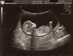
The baby will mostly be visible at about 7 weeks’ gestation; this is the period when it is easy to see baby’s heartbeat. In early pregnancy, detecting the heartbeat of a baby is almost impossible and proves to be a challenge for most doctors since you cannot conclusively determine if the baby is alive or not. If that is the case, then you may be told to return for another scan later. When you return for the scan, the doctor will be looking at the changes and differences in the pregnancy sac size as well as any development in the baby alongside heartbeat.
In some instances, you may have to go for several scans before your doctor detects the baby’s state or how the baby is developing. This uncertainty proves to be very difficult to deal with for some women.
What Exactly Can You See on Ultrasound?
When the doctor uses the ultrasound, some features of your baby appear on the screen and here is exactly what you can see.
1. Location of the Baby
When a 6-week pregnancy ultrasound is performed, it can easily determine the baby’s location, ascertaining that the baby is in the right location. It can also determine an ectopic pregnancy, depending on the patterns of blood flow that are seen on ultrasound.
2. Baby’s Heartbeat
Mostly, fetal heart beat will be detected at 6-week ultrasound. Normally at this time, the heart beat rate will be about 90 to 110 beats every minute. When the heart beat is detected at this time, it automatically means that the pregnancy will actually continue to the end and it will not end in a miscarriage; however, this is not a guarantee. If the heart beat cannot be detected at this time, the woman will be probably advised to come back again later, since if it not detectable at 6 weeks, it could be noticeable at 7 weeks or even after that.
3. Fetal Pole
This is the basic embryo shape which is normally seen around 6 weeks via the sonogram. It is bean-shaped and the doctor is able to determine its inclination, such as the rump and head ends of the baby. The fetus pole is very essential in measuring the length and size of embryo.
4. Yolk Sac and Chorionic Sac
Chorionic sac which is popularly known as the gestation sac is basically the fluid sac that embeds the fetus all throughout the pregnancy period. The yolk sac is within the chorionic sac and functions to provide the essential embryo nourishment. This is just for the time before the placenta develops. The yolk sac should be visible at this time as well as the chorionic sac.
What If the Baby Is Not Visible on Ultrasound?
The doctor will not only rely on the heartbeat, but they will most probably be concerned with the gestational sac, too. They will go ahead to measure the size of the embryo, which will help in determining the age of the baby. In case the baby is smaller than normal, then your due date might be off. Normally, the due date is basically an estimate which is based on the start of your last menstrual cycle. In addition, if an ultrasound is performed earlier than 6 week, it may not yield tangible results. If the baby is smaller, subsequent ultrasound checks may be required. And most importantly, if the baby’s heart beat is not visible after 6 weeks, then probably you may be in for an ectopic pregnancy or a miscarriage. However, this needs to be discussed with your doctor, since there could be other lab tests that may be essential before coming to a final conclusion.
Here is a fantastic video showing the stages of baby growth from 5 to 9 weeks in 3d. Check it out:
Other Types of Ultrasound That Can Be Used To See Your Baby
There are other types of ultrasound that can be used for this function as well.
|
Types of Ultrasound |
How It Works |
|
Doppler Imaging |
It measures the blood flow characteristics in your baby’s body. It is very essential when the mother has elevated levels of blood pressure or any deviation of blood pressure flow from the norm. You can have both tests at the same time since most of the ultrasound machines have the Doppler machines too. |
|
Trans-vaginal Ultrasound |
With this method, a transducer is usually placed in the vagina instead of moving it on the belly. It provides a better imagery especially in early pregnancy since the uterus is still small and near the vagina. |
|
Fetal Echocardiography |
It majorly detects any heart defects and gives a more defined image of your baby’s heart. |
|
Three-dimensional Ultrasound |
It gives more pronounced images or pictures of your baby. They look more life-like and give the examiners an easy task of evaluating your baby. |
|
Four-dimensional Ultrasound |
It is a four-dimensional ultrasound that mainly records your baby movements in the womb. |