An ultrasound can be exciting for the parents! It is a picture that is formed by using sound waves. In order to create the picture, the person doing the scan spreads a conductive gel on the mother’s belly, then glides a small handheld scanner over the gel. The gel allows sound waves to be passed through the uterus, and then they bounce back, creating an image. The ultrasound doesn’t hurt the mother or the baby, and can offer an exciting glimpse into the womb. A vaginal ultrasound could also be done, based on how many weeks pregnant you are. For those who are going to get an early ultrasound, you may wonder what happens at 8 week ultrasound and how your baby is developing at this stage.
Why May You Need to Do an Ultrasound at 8 Weeks?
This ultrasound is often a great moment for parents. It is the most common time for women to have the first ultrasound, just to make sure that everything is okay. Some ultrasounds might be performed for other reasons; here are a few of them:
- A “dating scan” can access gestational age
- If a mother is bleeding, the ultrasound can help determine the cause
- An ultrasound can confirm the presence of multiple pregnancies
- This ultrasound can check the size of the embryo, and confirm that there is a heartbeat
- An ultrasound can determine the health of the ovaries and fallopian tubes
- An ultrasound can rule out an ectopic pregnancy or other problems
What Happens at the 8 Week Ultrasound?
How it is done:
Sometimes the ultrasound is done with the wand across your belly, as explained above. But that might make things more difficult to see. In that case, a vaginal probe can be used to conduct the ultrasound. This is called a transvaginal ultrasound. It is performed with a small wand that is placed in the vagina and pressed against the cervix in order to get a picture from that direction. Just as with the other method, the ultrasound is not dangerous and does not hurt, though you might feel some pressure. Most women find that the pressure is barely noticeable, especially when they get to see their baby on the screen.
What you can see:
This first ultrasound gives you a great deal of information, including how the umbilical cord is functioning, the size of the placenta, the size of the embryo and the heart rate. You might also be able to tell if you have multiple babies in there. The ultrasound can confirm that everything is healthy and progressing as it should be.
After this ultrasound, others might be scheduled to ensure that the baby is growing as it should be. Some women prefer the transvaginal ultrasound because they are not required to have a full bladder for the scan to work. With the scan over the belly, the doctor might ask a woman to have a full bladder in order to “lift” the uterus up a bit. Fortunately, the later the pregnancy progresses, the less likely the doctor will be to want you to have a full bladder at the time of the ultrasound.
The following video shows how your baby looks like at 8 week ultrasound:
How Is Your Baby Developing at Week 8 – Gestational Age/Fetal Age 6 Weeks
At this point everything that an adult human has is now present in the embryo. This means that it is no longer an embryo and can be called a fetus. A strong fetal heartbeat should be clear on the ultrasound, and the heart rate should reach between 140 and 170 beats per minute by the 9th week of gestation.
Those who do not have a strong heartbeat will need to have a follow-up ultrasound to confirm the diagnosis because the fetus may not be viable. In this case, you will have two options: to miscarry naturally when the time comes, or to undergo a procedure called dilation and curettage, or D&C. About half of women who go through this kind of early pregnancy loss opt to miscarry naturally, while the other half opt for the D&C.
At this point, the baby already has tiny arms and legs, as well as buds where the hands and feet will be. It’s about 18 mm in length, the face is starting to take shape, and the internal organs are forming. The heart is already beating. There is even a tiny mouth, nostrils, tooth buds, and the beginning of eyes. The baby is moving quite a bit already, though mom can’t feel it yet.
Pictures of 8 Week Ultrasound
It is the first good look you will get of your baby! Here’s what to expect.
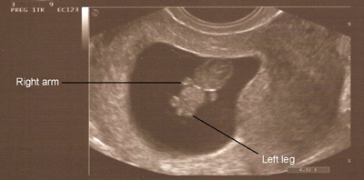
Picture 1: As you can see from this picture, the baby’s body is actually starting to look like a human. You can see little legs and arms, the head (which is much bigger than the body) and the space in which your baby is floating around.
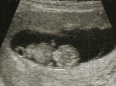
Picture 2: This is a more close up picture of the baby form a different angle. This view shows a much closer representation of the little body, including that round head and belly.
More Notes on Your hCG Level at 8 Week Pregnancy
In a normal pregnancy, hCG levels are a marker of how things are progressing. A good hCG level will peak at 8 to 12 weeks of pregnancy, and then slowly decline until it reaches a lower level. When a woman had bloodwork done and the results of the hCG test are questionable, an ultrasound can help the doctor decide if the pregnancy is viable or not. After five to six weeks of pregnancy, an ultrasound is considered more accurate than hCG levels in determining how viable the pregnancy really is.